Host defense is present in many forms. Overall, the Immune Response (IR) can be divided into two major classifications; humoral and cell-mediated. While these responses are not mutually exclusive, they provide distinctly different avenues for dealing with pathogenic organisms or altered host cells. These different responses will be discussed in more detail later.
Some of these responses are specific, others are non-specific. This page will introduce host defense mechanisms by defining some commonly used terms and describing the specific cells and tissues involved in these immune responses.
DEFINITIONS:
Antigen (Ag): A molecule which elicits a specific immune response when introduced into an animal. More specifically, antigenic (immunogenic) substances are:
- Generally large molecules (>10,000 daltons in molecular weight),
- Structurally complex (proteins are usually very antigenic),
- Accessible (the immune system must be able to contact the molecule), and
- Foreign (not recognizable as "self").
Antibody (Ab): A glycoprotein produced in response to an antigen that is specific for the antigen and binds to it via non-covalent interactions. The term "immunoglobulin" is often used interchangeably with "antibody". We will use the term "immunoglobulin" to describe
any antibody, regardless of specificity, and the term "antibody" to describe an
antigen-specific "immunoglobulin". Immunoglobulins (Igs) come in different forms (IgA, IgD, IgE, IgG, IgM) that reflect their structure.
More information can be found here.
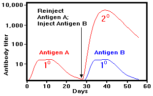
Antibody kinetics: The figure illustrates the production of antibody in response to antigenic substances. In this figure, an animal was injected with Antigen A at day 0. Antigen A invokes a primary response beginning about day 4, as indicated by a rise in the specific antibody titer (titer = measure of the amount of antibody in the animal's serum per unit volume). Initially, this antibody is mostly IgM (and some IgG). After a peak titer between days 7 and 10, the response decreases rapidly. If the animal is then reinjected with Antigen A at day 28, the production of antibody begins almost immediately and reaches a level 1000-fold greater that that seen in the primary response. This is known as the secondary response and the principal antibody produced is IgG. If a second antigen (Antigen B) is also injected at the same time as the reinjection of Antigen A, however, only a primary response to Antigen B is observed. These results demonstrate that:
- The immune response is specific.
- The immune response has memory.
Clonal selection hypothesis (Jerne and Burnet): The clonal selection hypothesis attempts to explain the findings described above by suggesting the following:
- Animals contain numerous cells called lymphocytes,
- Each lymphocyte is responsive to a particular antigen by virtue of specific
surface receptor molecules,
- Upon contacting its appropriate antigen, the lymphocyte is stimulated to proliferate (clonal expansion) and differentiate,
- The expanded clone is responsible for the secondary response (more cells to respond) while the differentiated ("effector") cells secrete antibody,
CELLS OF THE IMMUNE RESPONSE
Immune responsive cells can be divided into five groups based on i) the presence of specific surface components and ii) function: B-cells (B lymphocytes), T-cells (T lymphocytes), Accessory cells (Macrophages and other antigen-presenting cells), Killer cells (NK and K cells), and Mast cells. Some of the properties of each group are listed below.
| Cell group |
Surface components |
Function |
| B-lymphocytes |
- Surface immunoglobulin (Ag recognition)
- Immunoglobulin Fc receptor
- Class II Major Histocompatability Complex (MHC) molecule (Ag presentation)
|
- Direct antigen recognition
- Differentiation into antibody-producing plasma cells
- Antigen presentation within Class II MHC
|
|
|
|
| T-lymphocytes |
- CD3 molecule
- T-cell receptor (TCR, Ag recognition)
|
- Involved in both humoral and cell-mediated responses
|
|
|
- Recognizes antigen presented within Class II MHC
- Promotes differentiation of B-cells and cytotoxic T-cells
- Activates macrophages
|
|
|
- Downregulates the activities of other cells
|
|
|
- Recognizes antigen presented within Class I MHC
- Kills cells expressing appropriate antigen
|
|
|
|
| Accessory cells |
|
- Phagocytosis and cell killing
|
|
- Immunoglobulin Fc receptor
- Complement component C3b receptor
- Class II MHC molecule
|
- Bind Fc portion of immunoglobulin (enhances phagocytosis)
- Bind complement component C3b (enhances phagocytosis)
- Antigen presentation within Class II MHC
- Secrete IL-1 (macrokine) promoting T-cell differentiation and proliferation
- Can be "activated" by T-cell lymphokines
|
|
|
- Antigen presentation within Class II MHC
|
- Polymorphonuclear cells (PMNs)
|
- Immunoglobulin Fc receptor
- Complement component C3b receptor
|
- Bind Fc portion of immunoglobulin (enhances phagocytosis)
- Bind complement component C3b (enhances phagocytosis)
|
|
|
|
| Killer cells |
|
|
|
|
- Kills variety of target cells (e.g. tumor cells, virus-infected cells, transplanted cells)
|
|
- Immunoglobulin Fc receptor
|
- Bind Fc portion of immunoglobulin
- Kills antibody-coated target cells (antibody-dependent cell-mediated cytotoxicity, ADCC)
|
|
|
|
| Mast cells |
- High affinity IgE Fc receptors
|
- Bind IgE and initiate allergic responses by release of histamine
|
LYMPHOID TISSUES
|
|
|
|
| Primary |
Secondary |
| (Responsible for maturation of Ag-reactive cells) |
(Sites for Ag contact and response) |
 |
|
|
|
Thymus
(T-cell maturation) |
Bone marrow |
Lymph nodes |
Spleen |
| (T-cell maturation) |
(B-cell maturation) |
(Expansion of lymphatic system, separate from blood circulation. Deep cortex harbors mostly T-cells, superficial cortex harbors mostly B-cells) |
(Similar to lymph nodes but part of blood circulation. Collects blood-borne Ags) |